Formation of Facial Structures: Using Light Microscopy to Study the Causes of Facial Clefting
Introduction

Formation of Facial Structures: Using Light Microscopy to Study the Causes of Facial Clefting

Researchers use mouse embryos to provide insights into mechanisms underlying mammalian craniofacial development.

Craniofacial development is a complex morphogenetic process that requires a precise interplay of multiple cell and tissue types to generate the frontonasal skeleton. Disruption of this process results in highly prevalent human birth defects such as cleft lip and palate.

Dr. Katherine Fantauzzo is an Assistant Professor in the Department of Craniofacial Biology at the University of Colorado Anschutz Medical Campus, USA. Her research uses widefield light microscopy and mouse embryos to provide insight into the mechanisms underlying mammalian craniofacial development and new therapeutic directions for the treatment of human craniofacial birth defects.

Dr. Fantauzzo (left) and her former graduate student, Dr. Brenna Dennison, examine a skeletal preparation of a mouse embryo.

Dr. Fantauzzo (left) and her former graduate student, Dr. Brenna Dennison, examine a skeletal preparation of a mouse embryo.

Searching for the Genes Responsible for Cleft Lip and Palate

Mutations in the genes encoding a family of receptor tyrosine kinases, the platelet-derived growth factor receptor (PDGFR), cause non-syndromic cleft lip and palate as well as syndromes characterized by facial dysmorphism.

Defects in craniofacial development, including cleft lip and palate, comprise one of the most prevalent birth defects in humans. By identifying the mechanisms by which PDGFRs regulate gene expression and cell activity, our findings have the potential to provide new therapeutic directions for the treatment of human craniofacial birth defects.  

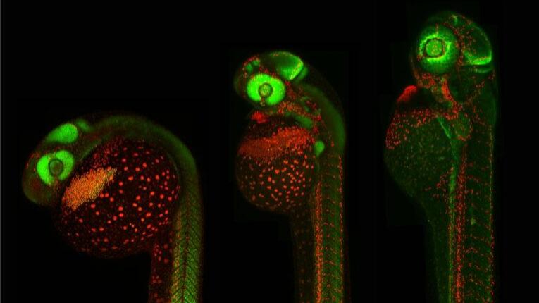
The Fantauzzo laboratory investigates the mechanism and function of the PDGFR family in the development of the mouse craniofacial skeleton.

Recent Discoveries

A Surprising Role for PDGFR Signaling in Regulating RNA Processing

Left: Whole-mount in situ hybridization analysis of Srsf3 expression in an E10.5 mouse embryo. Imaged using ZEISS Stemi 508 stereo microscope. Right: GFP-positive neural crest cells (green) and nuclei (blue) in a lateral whole mount fluorescence image of an E9.5 mouse embryo. Imaged using ZEISS Axio Observer 7 microscope.

Left: Whole-mount in situ hybridization analysis of Srsf3 expression in an E10.5 mouse embryo. Imaged using ZEISS Stemi 508 stereo microscope. Right: GFP-positive neural crest cells (green) and nuclei (blue) in a lateral whole mount fluorescence image of an E9.5 mouse embryo. Imaged using ZEISS Axio Observer 7 microscope.

Left: Whole-mount in situ hybridization analysis of Srsf3 expression in an E10.5 mouse embryo. Imaged using ZEISS Stemi 508 stereo microscope. Right: GFP-positive neural crest cells (green) and nuclei (blue) in a lateral whole mount fluorescence image of an E9.5 mouse embryo. Imaged using ZEISS Axio Observer 7 microscope.

A Surprising Role for PDGFR Signaling in Regulating RNA Processing

In B. Dennison et al., the team found that PDGFRα signaling regulates the expression of genes involved in palatal shelf morphogenesis through alternative RNA splicing. Phosphorylation of RNA-binding proteins downstream of this pathway were identified as a mechanism that contributes to this response. These findings further revealed that the RNA-binding protein Srsf3 is a critical regulator of craniofacial development in the mouse, as embryos with ablation of Srsf3 exhibit midline facial clefting. These results highlight a novel, and surprising, role for PDGFR signaling in regulating RNA processing.

Dr. Katherine Fantauzzo, Assistant Professor in the Department of Craniofacial Biology at the University of Colorado Anschutz Medical Campus, USA

Our ZEISS Axio Observer 7 microscope, is able to image the craniofacial region of mid-gestation mouse embryos in a single field of view at very high resolution.

Dr. Katherine Fantauzzo

Assistant Professor in the Department of Craniofacial Biology at the University of Colorado Anschutz Medical Campus, USA

Sox10-positive neural crest cells (magenta) as detected by immunofluorescence staining of a section of cranial neural folds from an E8.0 mouse embryo. Nuclei were stained with DAPI (blue). Imaged with ZEISS Axio Observer 7 with Apotome.

Sox10-positive neural crest cells (magenta) as detected by immunofluorescence staining of a section of cranial neural folds from an E8.0 mouse embryo. Nuclei were stained with DAPI (blue). Imaged with ZEISS Axio Observer 7 with Apotome.

Sox10-positive neural crest cells (magenta) as detected by immunofluorescence staining of a section of cranial neural folds from an E8.0 mouse embryo. Nuclei were stained with DAPI (blue). Imaged with ZEISS Axio Observer 7 with Apotome.

Microscopy to Explore Mouse Embryos, Craniofacial Tissues and Cells

The Fantauzzo lab uses microscopy to dissect mouse embryos and image these embryos as well as particular craniofacial tissues and individual cells. They do this using both brightfield and fluorescence microscopy.

In their recent publication, they used microscopy to assess the expression of Srsf3 during development and to determine the craniofacial phenotypes of mouse embryos upon Srsf3 ablation.

Journal Cover Feature Image

Dr. Brenna Dennison and Fantauzzo's recent work was published in Development. Their image of the cranial neural folds and first pharyngeal arches of a mouse embryo, acquired with a ZEISS Axio Observer 7 microscope with Apotome, was featured on the journal cover.

Image Selected for the Front Cover

Dr. Brenna Dennison and Fantauzzo's recent work was published in Development. Their image of the cranial neural folds and first pharyngeal arches of a mouse embryo, acquired with a ZEISS Axio Observer 7 microscope with Apotome, was featured on the journal cover.


Share this article