

Using Zebrafish to Understand How Blood and Lymphatic Vessels Develop
Zebrafish is an excellent animal model for biomedical research as it has a highly conserved genetic control of development and disease.
Dr. Kazuhide Shaun Okuda from the Peter MacCallum Cancer Centre in Victoria, Australia, uses ZEISS confocal microscopes to image zebrafish vessel development in real time. He is the winner of the LMA/VIA 2021 Image Competition of the Australian Microscopy & Microanalysis Society.


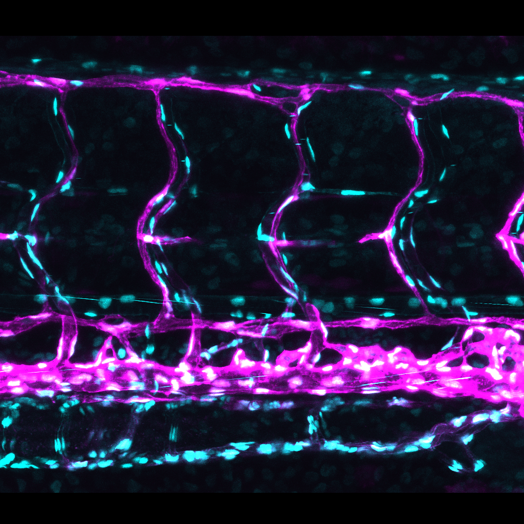
The above image displaying ribosomal biogenesis and blood vessels in developing zebrafish embryos was awarded first prize in the life sciences category of the LMA/VIA 2021 Image Competition (sponsored by ZEISS) of the Australian Microscopy & Microanalysis Society. Dr. Kazuhide Shaun Okuda, Postdoctoral Fellow at the Hogan laboratory, Peter MacCallum Cancer Centre in Victoria, Australia, took this image with a ZEISS confocal microscope at the Centre for Advanced Histology & Microscopy (CAHM).
As a child, whenever his mother took Dr. Okuda to the morning wet market in Malaysia, he would go straight to the fish vendor to marvel at all the different fish. He always wanted a fish but his mother would not allow it as he didn’t have a particularly good track record of keeping pets. Now he is surrounded by thousands of fish as he uses the zebrafish model to study how blood and lymphatic vessels develop. His expertise includes vascular biology, disease modelling, imaging and drug discovery.
We spoke to Dr. Okuda about his love of fish and latest discoveries:
Why zebrafish?
Zebrafish is an excellent animal model for biomedical research as it has a highly conserved genetic control of development and disease. The most important advantage of the zebrafish model is its optical transparency, allowing researchers like me to image blood and lymphatic vessel development in real time. This is still very challenging in optically dense mammalian models.
I can spend hours looking at these colorful vessels under a microscope without getting bored because they are so beautiful!


Cerebral vessels in zebrafish


Imaged with ZEISS confocal microscopy. Courtesy of Okuda Kazuhide Shaun.
Tell us about your research.
One research focus of mine is to generate and characterize genetically modified zebrafish (zebrafish transgenics) that enable visualization of the dynamic activity of key signaling pathways for blood/lymphatic development (recently published here). These zebrafish transgenics could also be utilized to understand molecular mechanisms of blood/lymphatic vessels in various pathological settings in real time. Another focus of my research is to uncover new lymphatic genes that may have functions in both developmental and pathological lymphangiogenesis. These genes could be a therapeutic target for various human diseases associated with excessive lymphangiogenesis, such as cancer and lymphatic malformation.


The zebrafish trunk vessel network

The Zebrafish Trunk Vessel Network
Imaged with ZEISS confocal microscopy. Courtesy of Okuda Kazuhide Shaun.
Would you like to highlight a specific discovery?
During my PhD, I contributed to the generation of the most lymphatic-specific fluorescent transgenic lines in zebrafish at the time (see publication). This allowed me to characterize previously hard-to-see lymphatic vessels in zebrafish. I was even given the privilege to name some of these lymphatic vessels! I am proud to say that these transgenic lines are currently one of the most utilized zebrafish models for live-imaging lymphangiogenesis globally.
How does confocal microscopy support your research?
I still remember the early days in my research career when all my experiments were done on fluorescent microscopes. While this gave us lots of valuable 2D data, the z-resolution information from confocal microscopy images allows us to visualize our data in 3D. This is particularly important for zebrafish research which relies heavily on live-imaging. Taking advantage of the large repertoire of zebrafish transgenics, I have used the confocal microscope to visualize multiple cell types in zebrafish including blood/lymphatic vessels, immune cells and neurons. I have also transplanted fluorescently labelled bacteria/cancer cells into zebrafish to image vascular responses in vivo. The resulting images and movies are not just aesthetically pleasing, but are packed with information that I can spend hours analyzing! It is all worth the effort when it’s published though!
Real-time visualization of blood vessel development and lumenization in zebrafish. Imaged using ZEISS confocal microscopy. Courtesy of Okuda Kazuhide Shaun.
What key findings do you present in your recent publication?
The winning image shows a zebrafish transgenic line we generated for our recent publication in the prestigeous journal Nature Cell Biology. The transgenic line enables real-time visualization of RNA helicase Ddx21 expression in zebrafish. A forward genetic screen conducted by my supervisor Professor Benjamin Hogan during his postdoc days in Utrecht, resulted in the identification of ddx21 zebrafish mutant, with reduced lymphatic vessel development. Surprisingly, blood vessel development was unaltered, showing that lymphatic vessels are selectively sensitive to loss of Ddx21. While Ddx21 is known for its function in ribosomal biogenesis, its role in vascular development had never been explored. Using this transgenic line, we showed that Ddx21 is expressed in zebrafish veins, which give rise to lymphatic vessels. Importantly, we showed that Vegfc/Vegfr3 signaling, essential for lymphangiogenesis, modulates Ddx21 level in endothelial cells. We are now interested in identifying inhibitors for Ddx21, which could be used to treat diseases like cancer and lymphatic malformation.


