Researchers Use C. elegans and Stereo Zoom Microscopy to Study Aging and Age-Related Diseases
Microscopy used to investigate disease models and the aging process
Aging is a universal trait among all organisms. From a public health perspective, with aging comes an increased risk of disease, including Alzheimer’s, cancer and metabolic disease/type II diabetes.
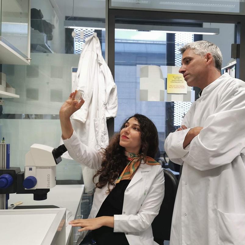
Dr. David Vilchez heads a lab at CECAD, (Cellular Stress Responses in Aging-Associated Diseases) in Germany. His team has recently published on impactful topics including new regulators of lifespan and links between healthy reproductive systems and age-related diseases. Their work incorporates innovative approaches including genetics in the model organism C. elegans and state-of-the-art proteomics. They use the ZEISS Axio Zoom.V16 microscope to visualize fluorescence proteins in C. elegans disease models or during the aging process in a living animal.
Human life expectancy has risen remarkably and the number of older people will continue to increase. We seek to understand the aging process and identify mechanisms to improve quality of life and prevent age-related diseases.
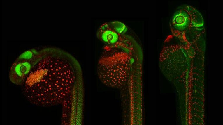
Left: Muscle actin cytoskeleton in young worms. Center: Muscle actin cytoskeleton in old worms, showing destabilization. Right: Preventing destabilization of muscle cytoskeleton in old worms by lowering the age-dysregulated high levels of EPS-8, a regulator of actin cytoskeleton. Imaged using ZEISS Axio Zoom.V16.
Left: Muscle actin cytoskeleton in young worms. Center: Muscle actin cytoskeleton in old worms, showing destabilization. Right: Preventing destabilization of muscle cytoskeleton in old worms by lowering the age-dysregulated high levels of EPS-8, a regulator of actin cytoskeleton. Imaged using ZEISS Axio Zoom.V16.
Imaging the Effects of Aging on Muscle Cytoskeleton
In S. Koyuncu et al., they show that the protein ubiquitin plays an important role in the regulation of the aging process. Through a comprehensive quantitative analysis of ubiquitin signatures during aging in C. elegans, theyfound site-specific information and defined quantitative changes in ubiquitin across all proteins in a cell during aging. In doing so, they discovered new regulators of lifespan and provided a comprehensive data set that helps to understand aging and longevity.
ZEISS Axio Zoom.V16 was used to image the cytoskeleton in filamentous actin with phalloidin, myosin heavy chain tagged to GFP and fluorescently tagged endogenous proteins of the intestine such as IFB-2 in C. elegans. These data were used to define how aging impairs the muscle cytoskeleton and the integrity of the intestine in these animals, and identified mechanisms to prevent these alterations.
Top: Mitochondria in healthy muscle of C. elegans. Bottom: Fragmented mitochondria in the muscle after induction of aggregation of proteins in the germline. Imaged using ZEISS Axio Zoom.V16.
Top: Mitochondria in healthy muscle of C. elegans. Bottom: Fragmented mitochondria in the muscle after induction of aggregation of proteins in the germline. Imaged using ZEISS Axio Zoom.V16.
Visualizing C. Elegans Age-Related Disease Models
In G. Calculli et al., the team found that a healthy reproductive system can prevent disease-related protein accumulation in distant tissues, such as neurons, and alteration of mitochondria. An imbalance of proteins, for example an aggregation of damaged proteins in brain cells, can lead to diseases like Alzheimer’s, Huntington’s disease or Amyotrophic Lateral Sclerosis (ALS).
Using C. elegans, they discovered that when the germline accumulates damaged protein aggregates, it releases specific signals (Wnt signaling) which in turn induce changes in mitochondria, leading to protein aggregation in other tissues such as muscle or neurons.
They used ZEISS Axio Zoom.V16 to image C. elegans germ line cells and mitochondrial morphology to look at protein aggregation during aging.
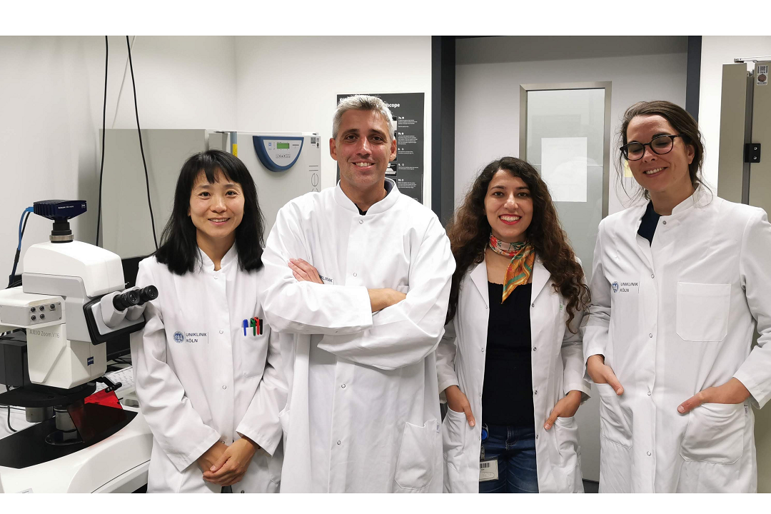
Members of the Vilchez Lab, from left to right: Dr. Hyun Ju Lee, Dr. David Vilchez, Dr. Seda Koyuncu and Dr. Rute Loureiro
What's Next
Dr. Vilchez and his team plan to apply their recent findings on ubiquitin modifications to identify novel mechanisms that can prevent age-related diseases that remain incurable, such as amyotrophic lateral sclerosis and Huntington’s disease.
They also seek to understand how the reproductive system communicates with neurons to regulate protein aggregation in these cells.